Dental X-Rays can be easy
Radiography is a fundamental part of modern veterinary dentistry, becoming an extension of the examination to allow a more thorough assessment of the oral cavity. This enables better diagnosis and results in improved treatment and outcomes for patients. Radiographs allow the veterinarian to see below the gum, identifying the presence of root remnants, periodontitis and resorptive lesions that may not be visible without radiographs. Interpretation of the radiographs relies on getting the best image possible.
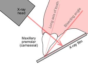
The basic principle of the bisecting angle technique is simple , however, it requires spatial awareness and can be a difficult technique to master in spite of training. At present, the recognised optimum technique for dental radiography in cats and dogs makes use of the size 2 film (using either DR or CR). This means that apart from one position (the lateral mandible view), all radiographs are taken using the bisecting angle technique. When performing dental radiographs on a regular basis the bisecting angle technique becomes second nature and is easy to grasp with minimal issues.. Nonetheless, the drawback of implementing this technique is that given the small size of the image plates, very often many retakes may be required in order to be able to achieve the required radiographic image, and it is likely that no interpretable image is obtained at all.
Thus the Magic 6 system was invented by iM3, with the aim of making it possible to achieve full-mouth radiographs on the majority of dogs and cats every time they have a scale and polish. The system is very simple and consists of bite plates, angle guides for dogs and cats and a vet size 5 plate for dogs.
The general principle is straightforward: the CR image plate is put into the bite protector, and the angle guide is placed in the edge of the bite protector in the centre. The angle guide will then provide the exact angle required (the correct bisecting angle), the distance from the CR plate and the centre of the CR Plate. For example, for a cat’s maxillary lateral view, the 30 degree angle guide and a size 2 film would be required. For dogs, a size 5 film and the positioners are longer as the distance to the generator needs to be greater; however, the general principle is the same.
The result of this new technique is that dental radiographs can be taken by every vet and nurse in the practice. This allows for consistent, good interpretable results.
Promoting full-mouth radiographs whilst the animal is having any dental treatment, such as a scale and polish, means at the end of the treatment the images can be reviewed and an appropriate treatment plan put in place, thereby providing excellent care and additional practice revenue.


